Wednesday, January 14, 2009

Medical Imaging for Early Detection of Diseases

One of the major benefits of medical imaging is the early detection of diseases. Breast cancers are spotted early on through mammogram. Even without having to touch a patient, doctors and medical practitioners are able to see a detailed view of a broken bone through x-ray. Changes in the internal organs and blood vessels are recognized through ultrasound. These are but few of the firsthand advantages of medical imaging. The whole human body is explored and new approaches for diseases are opened for treatment especially at their fundamental stages.

Mammography, CT Scans and Magnetic Resonance Imaging (MRI) are some of the most common medical imaging methods readily available to patients nowadays. These methods are routinely used throughout hospitals and health laboratories to detect cancer, heart diseases and other internal body complications. More complicated and expensive methods are electron microscopy, fluoroscopy, nuclear medicine, photo acoustic imaging, positron emission tomography (PET), projection radiography, tomography and ultrasound.

Tuesday, January 13, 2009

Medical Imaging for Cancer Detection

The January 16, 2007 issue of the Proceedings of the National Academy of Sciences published a study by researchers at Mount Sinai Hospital in New York that uses molecular MRI to gain insight into the correlation between inflammation and heart disease. Researchers developed a synthetic material, gadolinium–diethyltriaminepentaacetic acid (DTPA) that is able to track down and attach to white blood cells imbedded in arterial walls. The DPTA allowed the MRI visualization of the white blood cells, providing the ability to actually count the number of cells and assess their stability. Researchers found a positive correlation between the number of white cells imbedded in the arterial walls and the likelihood of subsequent heart attack. The initial research was conducted on mice. Further research will be conducted on larger animals and if successful, the research will move to human clinical trials.

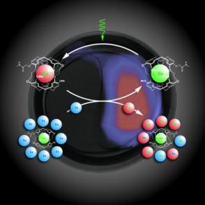
The search for better, more efficient and more specific medical imaging “tagging” media is the hottest new area of research in molecular magnetic resonance medical imaging. Recently, researchers with the U.S. Department of Energy's Lawrence Berkeley National Laboratory and the University of California at Berkeley have reported on research involving a new medical imaging technique for MRI that can detect molecules 10,000 times lower concentrations than conventional MRI techniques. The method, called HYPER-CEST, for hyperpolarized xenon chemical exchange saturation transfer, hyperpolarizes atoms with laser light to enhance their MRI signal, and then places the atoms into a nano-scale cage biosensor which is made specifically for a particular protein target. This medical imaging method is expected to be particularly useful in detecting cancer cells at the very earliest stages of cancer presence.

Sunday, January 11, 2009

What is Molecular Magnetic Resonance Imaging?


Magnetic resonance medical imaging, based on the principles of nuclear magnetic resonance, produces an image of the NMR signal in a thin slice through the human body. Images taken sequentially build a three dimensional picture of anatomical structures.Magnetic resonance medical imaging is the diagnostic tool of choice for visualizing the brain and spinal cord as well as evaluating soft tissue.

Molecular magnetic resonance medical imaging brings the level of visualization and analysis to the cellular and molecular level. At this level, it is possible to track and evaluate cellular functions that can provide never-before-available insight into the nature of the disease process. For example, there has long been an established connection between inflammation and heart disease. However, the medical imaging tools to measure inflammation related to the heart have simply not been available at a fine enough level of measurement to fully explore the connection.